Cavity-causing microbes can form superorganisms that can 'crawl'
Cavity-causing microbes can form superorganisms that can 'crawl'
Real-time microscopy allowed researchers to track the movement and behavior of a group of fungi and bacteria in saliva of children with severe dental caries. The interspecific group took on new functions and caused more severe decay than either species alone. (Image: Penn Dental Medicine)
A cross-kingdom partnership between bacteria and fungi may result in the two merging to form a "superorganism" with unusual strength and resilience. It may sound like science fiction, but these microbial groupings are an integral part of the here and now.
Found in the saliva of toddlers with severe childhood tooth decay, these assemblies can effectively colonize teeth. They were stickier, more resistant to antimicrobials and harder to remove from teeth than bacteria or fungi alone, according to the research team led by scientists from the University of Pennsylvania School of Dentistry. p>
Furthermore, the assemblies unexpectedly grow "limbs" that cause them to "walk" and "jump" to spread rapidly across the tooth surface, although each microbe itself is immobile, the team reported in the journal Proceedings of the National Academy of Sciences.
"It started with a very simple, almost accidental discovery by examining saliva samples from toddlers who develop aggressive tooth decay," says Hyun (Michel) Koo, professor at Penn Dental Medicine and co-author matching on paper. “Looking under the microscope, we noticed that bacteria and fungi were forming these assemblies and developing movements that we never thought they would possess: 'walking-like' and 'jumping-like' mobility. They have a lot of what we call “emerging features” that bring new benefits to this assembly that they couldn't achieve on their own. It's almost like a new organism, a superorganism, with new functions."
Better (or worse) together
In the past, Koo's lab has focused on dental biofilm, or plaque, present in children with severe tooth decay, finding that bacteria (Streptococcus mutans) and fungus (Candida albicans)—contributes to the disease. Cavities, commonly referred to as cavities, occur when sugars in the diet linger to feed bacteria and fungi in the mouth, resulting in acid-producing plaque that destroys enamel.
The new set of discoveries came when Zhi Ren, a postdoctoral fellow in Koo's group, used microscopy that allows scientists to visualize the behavior of living microbes in real time. The technique "opens up new possibilities for studying the dynamics of complex biological processes," says Ren, first author of the paper and a member of the first cohort of the NIDCR T90R90 postgraduate training program at Penn's Center for Innovation & Precision Dentistry.
After seeing the bacterial-fungal clusters in saliva samples, Ren, Koo and their colleagues were curious about how the clusters might behave once attached to a tooth surface. Thus began a series of experiments using live microscopy in real time to observe the process of attachment and eventual growth.
As the assembly grew, it also began to move, the researchers found. Fungal projections (in blue) propelled bacteria (in green) along the surface of a tooth in a jumping motion. (Image: Penn Dental Medicine)
 Real-time microscopy allowed researchers to track the movement and behavior of a group of fungi and bacteria in saliva of children with severe dental caries. The interspecific group took on new functions and caused more severe decay than either species alone. (Image: Penn Dental Medicine)
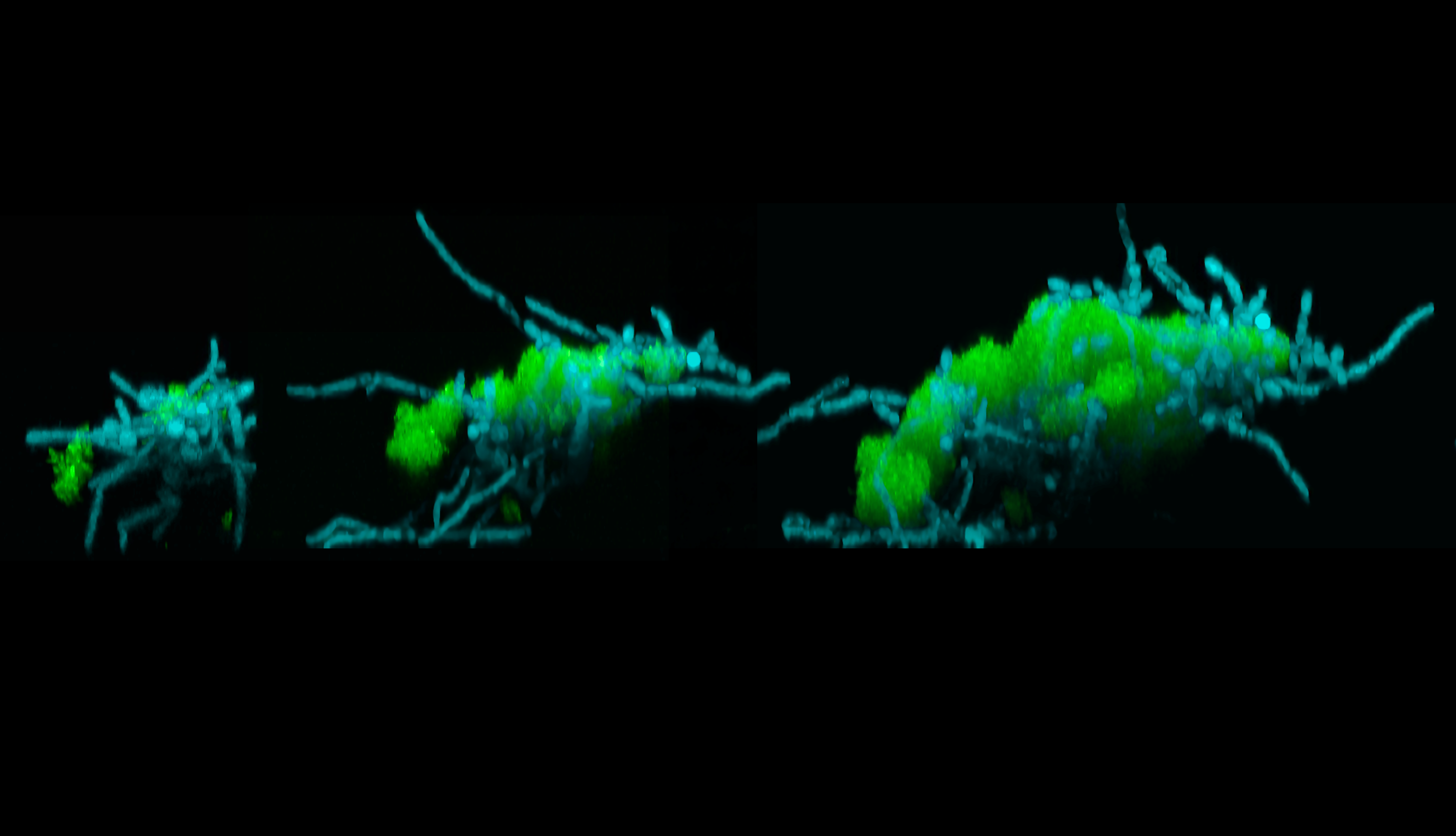 As the assembly grew, it also began to move, the researchers found. Fungal projections (in blue) propelled bacteria (in green) along the surface of a tooth in a jumping motion. (Image: Penn Dental Medicine)